Se habla español
Retinal disease covers a broad group of conditions that can subtly reduce sight or cause sudden, serious vision loss. At Next Level Retina in Oak Brook, Illinois, Dr. Neel Lamba, MD, MBA — a board-certified ophthalmologist with fellowship training in vitreoretinal disease — evaluates and treats the full spectrum of retinal disorders. Our goal is clear: identify problems early, preserve function where possible, and guide patients through treatment choices with clarity and compassion.
The retina is the light-sensitive layer at the back of the eye that converts incoming light into electrical signals. These signals travel along the optic nerve to the brain and are interpreted as images. Because the retina performs this essential conversion, even small disruptions can have an outsized effect on visual clarity, color perception, and the ability to read or recognize faces.
Different regions of the retina serve different visual tasks. The macula, for example, supports sharp central vision and fine detail, while the peripheral retina helps with motion detection and spatial awareness. Understanding these functional zones helps clinicians pinpoint the source of symptoms and tailor diagnostic testing and treatments appropriately.
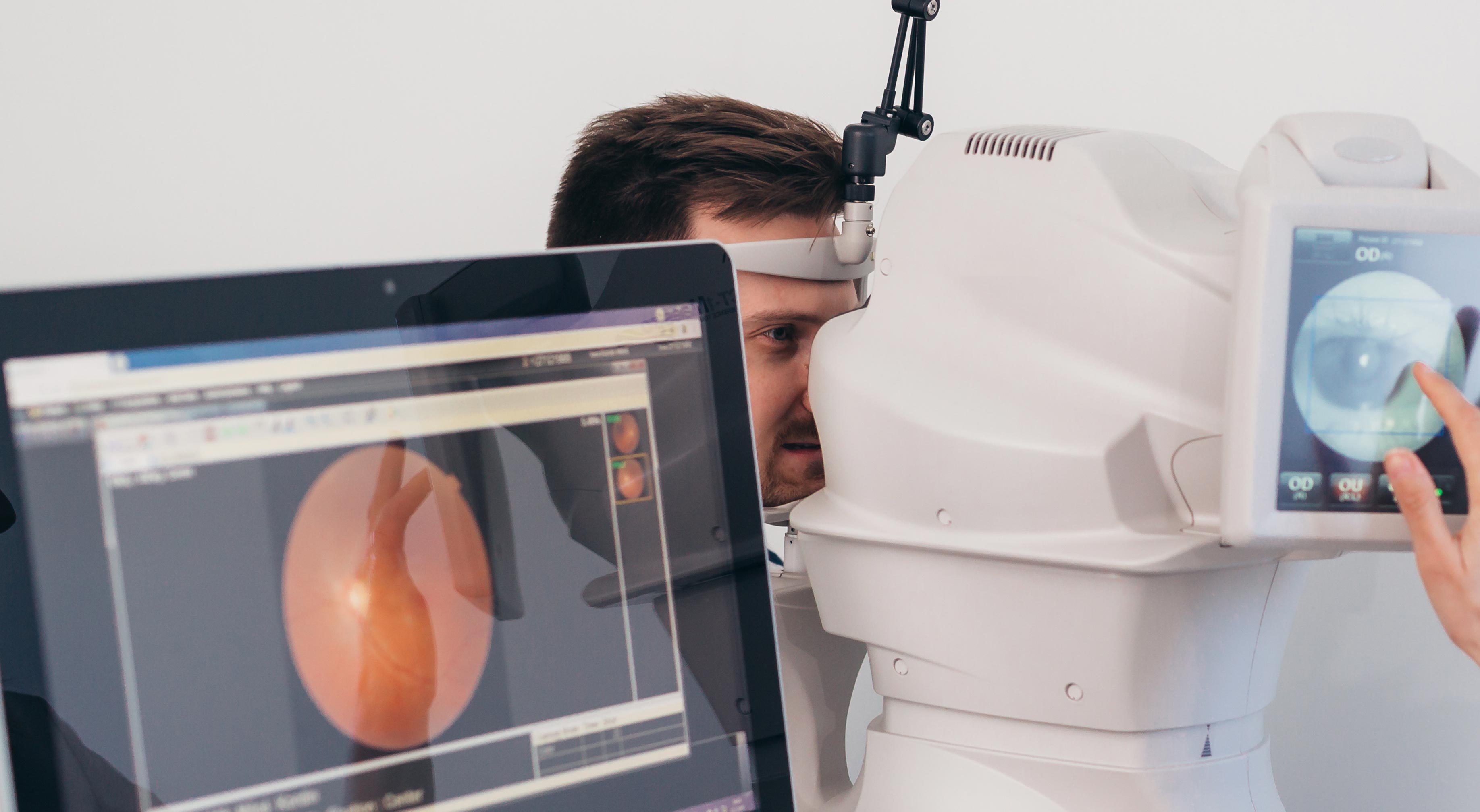
Advances in retinal imaging and surgical techniques have improved clinicians’ ability to detect disease earlier and intervene more effectively. Tests such as optical coherence tomography (OCT), wide-field photography, and fluorescein angiography allow precise visualization of retinal structure and blood flow, which supports accurate diagnosis and individualized care planning.
Retinal conditions can present in many ways, and early signs are not always dramatic. Patients often notice changes in familiar tasks — reading small print becomes harder, straight lines appear wavy, or dark spots drift through the field of vision. These symptoms can progress slowly or come on suddenly, and any persistent change in vision should prompt evaluation.
Common warning signs that suggest a retinal problem include new or increasing floaters, flashes of light, sudden blurring, a dark curtain moving across part of the vision, or trouble seeing in dim light. Symptoms that affect only one eye or that appear abruptly warrant urgent assessment to rule out conditions like retinal detachment or vascular occlusion.
Because some retinal diseases progress with few early symptoms, routine dilated retinal exams are an important preventive tool — particularly for people with systemic risk factors. Early detection not only expands treatment options but can also preserve independence and quality of life over the long term.
Several health and lifestyle factors raise the likelihood of retinal disease. While some risks — such as age and inherited genetics — cannot be changed, many patients can reduce their risk through medical management and healthy behaviors. A thorough health history and collaborative care with your primary physician help clinicians identify which preventive measures are most relevant.
The risk of many retinal conditions climbs with age. Age-related macular degeneration (AMD) and changes related to long-standing systemic disease become more common as the retina ages, making regular monitoring essential for older adults.
Certain retinal disorders have genetic roots. A family history of conditions like retinitis pigmentosa or familial macular dystrophies can suggest an inherited risk that merits earlier or more frequent screening, and genetic counseling may be appropriate for some families.
Diabetes, hypertension, and some autoimmune conditions can directly affect retinal blood vessels and tissue. Effective control of blood sugar, blood pressure, and systemic inflammation reduces the likelihood of progressive retinal damage and complements eye-specific treatments.
Direct injury to the eye can cause tears, hemorrhage, or detachment of the retina. Any eye trauma deserves prompt specialist evaluation, even if initial symptoms seem minor, because early repair can prevent irreversible vision loss.
Long-term exposure to ultraviolet light, certain medications, and toxic agents can contribute to retinal stress over time. Protective eyewear, careful medication review, and routine eye examinations help mitigate these risks when possible.
Treatment for retinal disease is highly individualized and depends on the diagnosis, severity, and patient goals. Modern therapy often uses a combination of pharmacologic, laser, and surgical techniques designed to stabilize vision, reduce symptoms, and address the underlying cause of damage.
Intravitreal injections of anti-VEGF medications are a cornerstone treatment for conditions that involve abnormal blood vessel growth or leakage, such as wet AMD and some forms of diabetic retinopathy. These injections reduce fluid and hemorrhage, often improving or stabilizing vision when given on a regular schedule determined by clinical response.
Laser photocoagulation remains a valuable option for sealing leaking vessels, treating localized areas of retinal ischemia, or creating adhesion around retinal tears. Laser procedures are performed in-office and can be an effective, targeted therapy in many cases.
When the retina requires mechanical repair — for example, to remove scar tissue, clear persistent vitreous hemorrhage, or reattach a detached retina — vitrectomy and related surgical techniques offer durable solutions. These procedures are performed by fellowship-trained vitreoretinal surgeons and are tailored to maximize anatomical and functional outcomes.
Steroid injections into the eye can control inflammation and reduce macular swelling when inflammation contributes to vision loss. They are an important adjunct for conditions that are not fully responsive to other therapies, and their use is guided by the expected benefits and potential side effects.
For a subset of patients with advanced, otherwise untreatable retinal degeneration, implantable devices and prosthetic technologies may offer partial restoration of visual function. These are specialized options that require careful evaluation to identify appropriate candidates.
Successful retinal care often extends beyond a single procedure or injection. Ongoing monitoring, timely retreatment when indicated, and coordination with primary care and other specialists form the backbone of sustained visual health. Personalized follow-up schedules help detect recurrence or progression early, when interventions are most effective.
Lifestyle choices and systemic disease control are important complements to eye-specific treatment. Smoking cessation, maintaining healthy blood sugar and blood pressure, wearing UV-protective eyewear, and attending regular dilated retinal exams all contribute to better outcomes. Patients and clinicians working together can often slow the course of disease and preserve meaningful vision.
When vision changes are permanent, low-vision rehabilitation and adaptive strategies can help patients maintain independence and quality of life. The practice coordinates with vision rehabilitation specialists and support services to ensure patients have access to the tools and training they need.
Retinal disease requires timely attention and an approach that combines expert diagnosis, evidence-based treatment, and thoughtful long-term planning. If you have noticed changes in your vision or have risk factors for retinal problems, Dr. Neel Lamba and the team at Next Level Retina are available to evaluate your condition and discuss tailored care options. Contact us for more information.
The retina is the light-sensitive tissue at the back of the eye that converts light into electrical signals the brain interprets as vision. Because this layer performs the retina's essential functions, even small disruptions can significantly affect visual clarity, color perception and central detail. Retinal disease refers to a broad group of disorders that damage retinal cells, blood vessels or supporting structures and can lead to gradual or sudden vision loss.
Conditions that fall under retinal disease include age-related macular degeneration, diabetic retinopathy, retinal tears and detachment, inherited dystrophies and vascular occlusions. Symptoms and progression vary widely by diagnosis, which is why a tailored diagnostic workup is essential. At Next Level Retina in Oak Brook, Illinois, specialists evaluate each case individually to define the diagnosis and outline a personalized plan of care.
Early warning signs of retinal problems can be subtle and are not always painful. Patients commonly report new floaters, intermittent flashes of light, blurred or distorted central vision and areas of missing or shadowed vision. A sensation like a curtain or veil moving across part of the visual field is particularly concerning and often indicates an urgent issue.
Because many retinal conditions progress without obvious early symptoms, routine dilated retinal exams help detect problems before they threaten sight. Even minor or temporary symptoms should not be dismissed, especially if they occur suddenly or affect only one eye. Rapid assessment increases the chances of successful treatment and better long-term outcomes.
Retinal disease arises from a mix of age-related degeneration, systemic vascular disease, genetic mutations, inflammation and mechanical injury. Age increases the risk for conditions such as macular degeneration while long-standing diabetes and hypertension damage retinal blood vessels and elevate the risk of bleeding, swelling and ischemia. Inherited conditions like retinitis pigmentosa are caused by gene variants that primarily affect photoreceptors and retinal metabolism.
Trauma and certain medications or toxic exposures can also precipitate retinal damage in susceptible individuals. A comprehensive medical history and coordination with primary care providers help identify modifiable risk factors and guide screening frequency. Genetic counseling may be recommended when an inherited condition is suspected to inform family planning and targeted surveillance.
Diagnosing retinal disease begins with a dilated eye exam and symptom review, allowing the specialist to assess retinal structure and function directly. Advanced imaging tools such as optical coherence tomography (OCT), wide-field retinal photography and fluorescein angiography provide high-resolution views of retinal layers, fluid accumulation and blood flow. These tests enable precise localization of pathology and support data-driven treatment decisions.
Functional testing, including visual acuity, Amsler grid assessment and visual field testing, complements imaging to measure how the disease affects daily tasks. In select cases, electroretinography or genetic testing may be used to clarify diagnoses or guide prognostic counseling. Together, these evaluations form the basis of a personalized care plan with scheduled follow-up to monitor response to therapy.
Treatment for retinal disease is individualized and may include medical, laser and surgical options aimed at preserving or restoring vision. Intravitreal injections of anti-VEGF agents and intraocular steroids reduce abnormal vessel growth and swelling in many vascular retinal disorders. Laser photocoagulation can seal leaking vessels or create adhesion around retinal tears, while newer laser techniques minimize collateral tissue damage.
When structural repair is needed, vitreoretinal surgery such as pars plana vitrectomy can remove scar tissue, clear hemorrhage and reattach the retina. The choice and timing of surgery depend on the specific condition, extent of damage and the patient's visual goals. Emerging technologies, including retinal implants for selected advanced degenerations, offer additional options for carefully selected patients.
Certain visual symptoms require immediate evaluation because they can signal sight-threatening conditions such as retinal detachment or central retinal artery occlusion. Seek urgent attention if you experience a sudden increase in floaters, new flashes of light, a dark curtain coming across the vision or abrupt, severe loss of vision in one eye. Timely assessment and intervention are critical to maximizing the chance of anatomical repair and visual recovery.
Even when symptoms resolve quickly, follow-up with a retinal specialist is important to document findings and monitor for progression. If you have risk factors such as recent eye surgery, trauma or known retinal tears, establish rapid-access pathways with your eye care team. Early communication between referring clinicians and the retinal specialist helps ensure prompt triage and coordinated care.
Diabetes is a leading cause of retinal disease and can produce a spectrum of changes collectively called diabetic retinopathy. High blood sugar damages small retinal blood vessels, leading to microaneurysms, hemorrhage, fluid accumulation in the macula and, in advanced stages, abnormal new vessel growth. Regular retinal screening for people with diabetes enables early detection of these changes before they impair daily activities.
Management combines systemic control of blood glucose and blood pressure with eye-specific treatments such as anti-VEGF injections, focal laser or surgery when indicated. Close collaboration between the retina specialist and the patient's primary care provider or endocrinologist optimizes overall risk reduction and treatment timing. Structured follow-up and patient education about symptom recognition are essential to prevent vision loss.
At an initial retinal consultation you can expect a detailed history, symptom review and a comprehensive dilated examination to evaluate retinal health. Imaging tests such as OCT and wide-field photography are commonly performed during the visit to document retinal structure and guide decision-making. The specialist will explain findings in plain language and discuss a personalized plan that balances risks, benefits and patient goals.
When appropriate, Dr. Neel Lamba will outline likely treatment paths, expected follow-up intervals and functional milestones to monitor progress. Staff will also provide instructions for urgent symptoms and available support services such as vision rehabilitation if needed. This collaborative approach ensures patients understand their condition and participate actively in long-term care.
Reducing long-term risk of retinal damage involves both medical management and lifestyle choices to protect retinal circulation and cellular health. Key measures include smoking cessation, strict control of blood sugar and blood pressure, regular exercise and wearing UV-protective eyewear when appropriate. Medication reviews with your clinician can identify agents that may affect retinal health and prompt alternatives when necessary.
Routine dilated retinal exams allow early detection and intervention, particularly for individuals with diabetes, a family history of retinal disease or advancing age. Adherence to scheduled follow-up and prompt reporting of new symptoms amplify the effectiveness of preventive efforts. A coordinated care team helps align systemic disease management with ocular surveillance to improve long-term outcomes.
When vision loss is permanent, a range of low-vision services can help people maintain independence and quality of life. Low-vision rehabilitation includes training with magnification devices, adaptive technologies, occupational therapy and strategies for daily living. Rehabilitation specialists assess functional needs and recommend tools and techniques to maximize remaining vision.
Next Level Retina coordinates care with vision rehabilitation providers, social services and assistive technology experts to create practical support plans for patients. Your retinal specialist can also provide referrals and documentation to access community resources, adaptive devices and vocational services when appropriate. Ongoing medical follow-up remains important to monitor for treatable complications and to adjust support as needs change.
Phone:
Fax: